Back to the Gastrointestinal Image Gallery
Collages
The Gastrointestinal Video Gallery
The Gastrolab Image Gallery
SLIDE SHOW: The Normal Gastrointestinal Channel
This free script provided by
JavaScript Kit
Larynx with the vocal cords clearly visible. Just click on the picture to get a magnification.
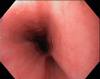
Oesophagus
Cardia
x
Gastric fundus, the "roof of the stomach" seen from below with the endoscope in an inverted position
A normal gastric body (the upper half of the stomach, corpus ventriculi)
Peristaltic wave in the gastric antrum
The gastric antrum and pylorus
A normal duodenal bulb seen through the pylorus
A normal duodenal bulb (bulbus duodeni)
A normal descending duodenum
Ileum terminale, the very last part of the small bowel
A normal ileocaecal valve (valvula Bauhini)
Appendix aperture in the caecum
The ileocaecal valve (valvula Bauhini), caecum and the first part of the ascending colon
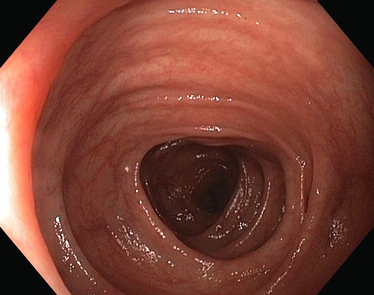
Colon transversum
Colon descendens
A normal sigmoid colon
A normal rectum
Ampulla recti
Linea dentata (also called linea anorectalis, anorectal junction, anocutaneous line) seen from the inside of the rectum with the endoscope in an inverted position
Anus
You might also be interested in:
A normal oesophagus
Hiatal Hernia from Below
A Normal Antrum and Pylorus
A Normal Descending Duodenum
A Normal Caecum
 A normal oesophagus
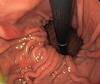 Hiatal Hernia from Below
 A Normal Antrum and Pylorus
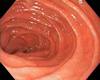 A Normal Descending Duodenum
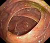 A Normal Caecum